

Tracking and Shape Segmentation of Atrial Septal Defects in 3D echocardiography
Coworkers: M. G. Linguraru, G. R. Marx, P. J. del Nido, R. D. Howe
Real-time cardiac ultrasound allows monitoring the heart motion during intracardiac beating heart procedures. Our application assists atrial septal defect (ASD) closure techniques using real-time 3D ultrasound guidance. One major image processing challenge is the low image quality, especially given the required processing of information at high frame rate. We present an optimized block flow technique, which combines the probability-based velocity computation for an entire block with cyclic template matching. We propose adapted similarity constraints both from frame to frame to conserve energy, and globally from a reference template to minimize errors. Computing velocity at the block level with an optimized scheme, our technique tracks ASD motion at a frequency of 60 frames/s on clinical 4D datasets. ASD tracking is also an important tool towards systematic clinical studies of the dynamic behavior of the intra-atrial communication. In particular, we show real-time tracking and preliminary segmentation results of the ASD shape and orientation as a function of time.
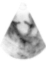
Ultrasound image of the heart.
ASD is the hole visible in the upper membrane of the picture.
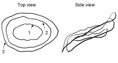

4D shape segmentation of the ASD.